1. Introduction – Where Did Life Begin?
Scientists widely believe that life originated in water. Some researchers think life may have started in small water pools with changing environmental conditions – not in the oceans. Hot springs are a good example of such environments.
In India, the hot springs of Puga Valley in Ladakh maintain very high temperatures (nearly at the boiling point of water) even in cold climates. These conditions are believed to be similar to early Earth, about 3.5 billion years ago.
The organisms living in these hot springs are mostly heat-loving bacteria called thermophiles, which are unicellular. Scientists from the Birbal Sahni Institute of Palaeosciences, Lucknow, found that calcium carbonate deposits formed rapidly around these springs. These deposits may have:
- Protected early organic molecules from harmful radiation
- Helped form the first protective membrane – the barrier that defines a cell
2. What is a Cell?
The cell is the basic structural and functional unit of all living organisms. Every living thing – from the tiniest bacterium to a giant tree or a human being – is made up of cells.
Types of organisms based on number of cells:
| Type | Meaning | Examples |
|---|---|---|
| Unicellular | Made of only one cell | Bacteria, yeast, amoeba |
| Multicellular | Made of millions of cells working together | Plants, fish, birds, humans |
Organisation in multicellular organisms:Cells → Tissues → Organs → Organ Systems
Example: Nasal pores + nasal cavity + trachea + lungs = Respiratory System
Even when cells are organised into tissues and organs, the cell remains the fundamental unit of structure and function.
3. How to Study Cells?
Cells are too small to be seen with the naked eye. The limit of resolution of the human eye is 0.1 mm – meaning two points closer than 0.1 mm cannot be seen as separate.
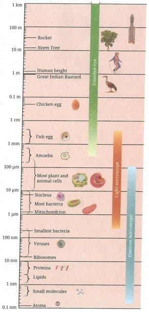 Size of the objects and its visibility through unaided to aided eye
Robert Hooke was the first person to observe a cell in 1665 using a self-designed microscope (200-300X magnification). He observed a thin slice of cork and saw small box-like compartments – he named them ‘cells’.
Types of microscopes:
| Microscope | What it does |
|---|---|
| Light Microscope | Uses visible light; used in school labs; magnifies using objective lens (10X, 40X) and eyepiece |
| Electron Microscope | Uses a beam of electrons instead of light; shows cell structure at the nanometre scale (1 nm = 0.000001 mm); much more powerful than a light microscope |
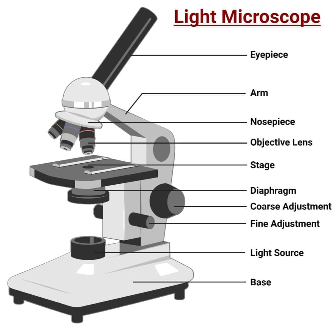 Light Microscope
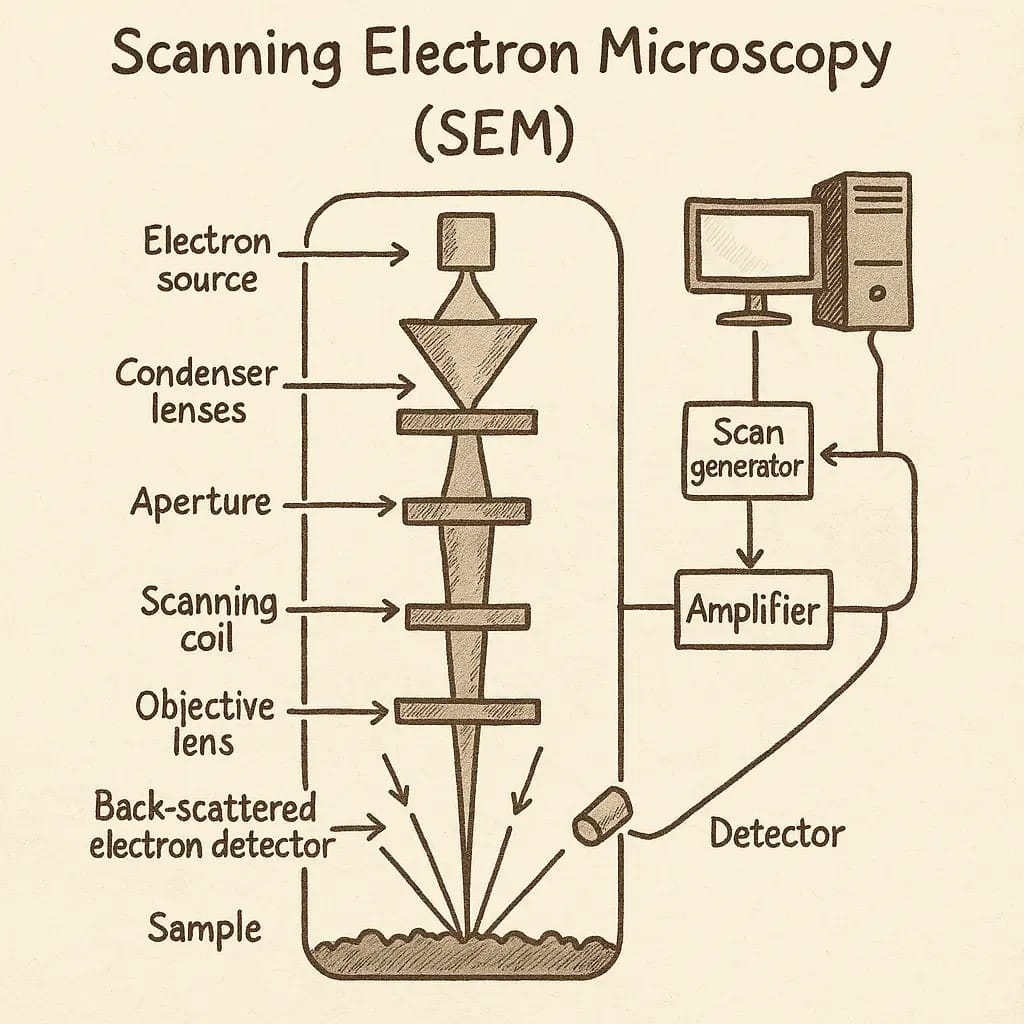 Electron Microscope
Three features that have improved over time:
- Resolution – measure of clarity (how clearly two close points can be seen)
- Contrast – difference in brightness between different parts of an object
- Magnification – how much larger the object appears
How to estimate the size of a cell (Activity 2.1):
Formula:
Estimated size of cell = Diameter of visible field (in µm) ÷ Number of cells along the diameter
Example: If diameter = 5 mm = 5000 µm, and 25 cells are seen along the diameter: Size of one cell = 5000 ÷ 25 = 200 µm
If eyepiece = 10X and objective = 10X → total magnification = 100X → the 200 µm cell appears 100 times larger.
Unit conversion: 1 mm = 1000 µm (micrometre)
4. Structure of a Cell
Every cell has a boundary – the cell membrane – through which substances move between the cell and its external environment. Even single-celled organisms exchange materials through the cell membrane.
4.1 Cell Membrane – The Universal Feature of a Cell
The cell membrane (also called plasma membrane) is a thin boundary that:
- Surrounds and protects the cell
- Defines the individuality of a cell
- Is selectively permeable – allows some substances to pass through while blocking others
Structure – Fluid Mosaic Model:The cell membrane is extremely thin – about 7 to 10 nanometres (nm) thick. It is made of lipids (fats) and proteins.
Structure of cell membrane
- It has a lipid bilayer – two layers of fat molecules with water-attracting heads pointing outward and water-repelling tails pointing inward
- Proteins are embedded in this bilayer and act like gatekeepers – controlling what enters and exits
- The molecules can move sideways, flip, and rotate → that is why it is called fluid
- The molecules are arranged like tiles in a mosaic → that is why it is called mosaic
Together this is the Fluid Mosaic Model.
4.2 Osmosis and Diffusion
Diffusion is the net movement of particles from an area of higher concentration to an area of lower concentration. This happens even without a membrane.
Osmosis is the diffusion of water specifically, through a selectively permeable membrane, from an area of more water (less solute / dilute) to an area of less water (more solute / concentrated), until concentrations become equal.
Simple way to remember: In osmosis, water moves from dilute solution → concentrated solution through a membrane.
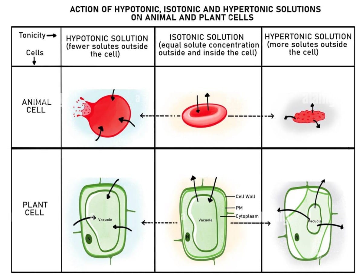
Types of solutions and their effect on a cell:
| Solution Type | What it means | Effect on cell |
|---|---|---|
| Isotonic | Solute concentration outside = inside | Cell stays the same – no change |
| Hypotonic | Solute concentration outside < inside | Water enters cell → cell swells |
| Hypertonic | Solute concentration outside > inside | Water leaves cell → cell shrinks |
Example from Activity 2.2 (Potato experiment):
Experimental set-up, and initial andfinal states of potato pieces in (a) plain water, and (b) 20 per cent salt solution
- Potato in plain water (hypotonic) → swells (water enters by osmosis)
- Potato in 20% salt solution (hypertonic) → shrinks (water leaves by osmosis)
In plants, water from soil enters root cells by the process of osmosis.
4.3 Cell Wall – The Outer Covering of Cells
Plants cannot move, so they need extra rigidity and support. That is why plant cells have an additional layer outside the cell membrane called the cell wall.
Key features of the cell wall:
- Present in plants, fungi, and bacteria – absent in animal cells
- Made primarily of cellulose (a carbohydrate made of many glucose units)
- Rigid but permeable – water and dissolved minerals can pass through
- Helps leaves and flowers stay firm, maintains shape, keeps plants upright
What happens when a plant cell loses water (osmosis)?
- Plant cells placed in concentrated sugar solution lose water → inner content shrinks
- But the outer boundary (cell wall) stays the same – it holds the shape
- The cell membrane pulls away from the cell wall – this is called plasmolysis
Animal cells (e.g., cheek cells) have no cell wall. When placed in concentrated solution, they simply shrink because there is no rigid wall to maintain shape.
Without a rigid cell wall, animal cells can change shape freely – this is actually useful. It allows animal cells and tissues to move and flex.
5. The Cell Interior – A Coordinated Working System
Every cell has three basic parts:
- Cell membrane – selectively permeable outer boundary
- Cytoplasm – semi-fluid, jelly-like substance that fills the cell
- Nucleus – the control centre of the cell
In eukaryotic cells, the cytoplasm also contains organelles – small specialised structures that each perform a specific job. Think of a cell as a tiny factory where each organelle is a department doing its own work.
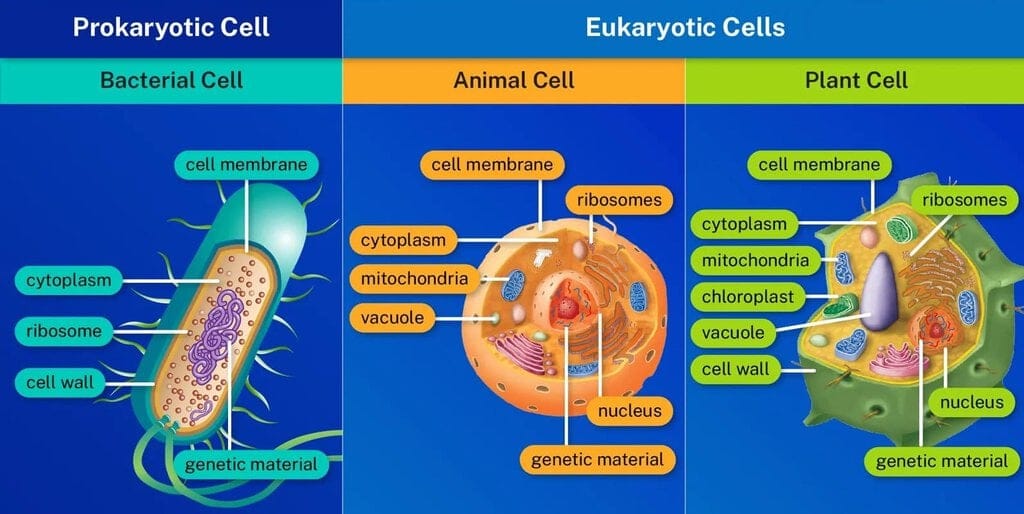
5.1 Prokaryotic vs Eukaryotic Cells
| Characteristics | Prokaryotic Cell | Eukaryotic Cell |
|---|---|---|
| Nucleus | No well-defined nucleus (only nucleoid region) | Well-defined nucleus with nuclear membrane |
| Diameter | 1 to 10 µm | 10 to 100 µm |
| Membrane-bound organelles | Absent | Present |
| Examples | Bacteria | Plant cells, animal cells, fungi |
Pro = primitive, karyon = nucleus → prokaryotic = primitive nucleusEu = true, karyon = nucleus → eukaryotic = true nucleus
In prokaryotic cells, most activities happen directly in the cytoplasm. Their DNA is present as a single circular molecule in a region called the nucleoid (not enclosed by a membrane).
5.2 Cell Organelles in Detail
A. Nucleus – House of Coded Instructions
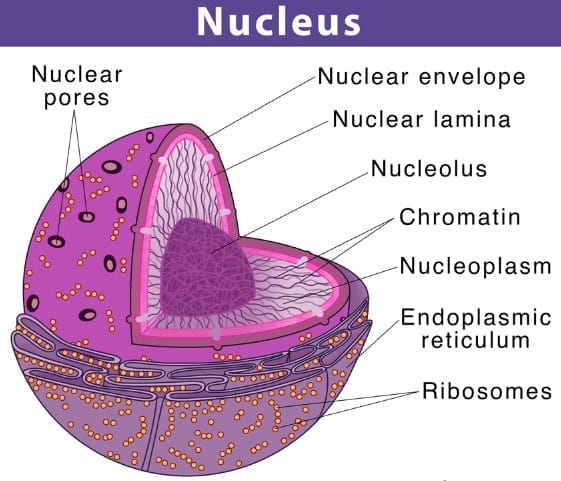
The nucleus is the control centre of the cell. It contains the genetic instructions for all cell activities.
Structure:
- Surrounded by a double-layered nuclear membrane with nuclear pores – allow transfer of material between nucleus and cytoplasm
- Contains the nucleolus – a dense round body inside the nucleus where ribosomal subunits are made
- Contains chromosomes – visible as rod-shaped structures only when the cell is about to divide
- Chromosomes are made of DNA + proteins
- DNA contains the genetic information. Functional segments of DNA are called genes
- When the cell is not dividing, DNA exists as chromatin – an entangled mass of thread-like material
- When the cell is about to divide, chromatin organises into chromosomes
Interesting fact: Mature Red Blood Cells (RBCs) in humans have no nucleus. The absence of a nucleus provides more space for haemoglobin, allowing more oxygen to be transported. Because they have no nucleus, RBCs cannot repair or divide themselves – their lifespan is only about 120 days.
In prokaryotic cells, DNA is present as a single circular molecule in the nucleoid region – not enclosed by any membrane.
B. Ribosomes – The Protein Factories
- Tiny structures found either freely in the cytoplasm or attached to the endoplasmic reticulum
- Function: Site of protein synthesis – they build proteins using instructions from DNA
- Present in both prokaryotic and eukaryotic cells
C. Endoplasmic Reticulum (ER) – Manufacturing Factory
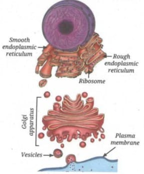
The ER is a large organelle that spreads like a network through the cytoplasm. It is continuous with the outer nuclear membrane.
Function: Synthesis and transport of proteins, fats (lipids), and some hormones
Two types:
| Type | Appearance | Function |
|---|---|---|
| Rough ER (RER) | Rough – has ribosomes on its surface | Protein synthesis and secretion (e.g., in gland cells like pancreatic cells) |
| Smooth ER (SER) | Smooth – no ribosomes | Synthesis and storage of fats (lipids) and hormones |
D. Golgi Apparatus – The Packaging and Shipping Centre
- Made of stacks of flattened, sac-like structures
- First observed in 1898 by Italian scientist Camillo Golgi in nerve cells of a barn owl
- Functionally linked to the ER and cell membrane
Function: Modifies, sorts, and packages proteins and/or lipids received from the ER into vesicles – small membrane-bound packets – for transport, secretion, or lysosome formation
Think of it as the post office of the cell – it receives, packages, and ships materials to the right destinations.
E. Lysosomes – The Clean-Up System
- Single membrane-bound sacs filled with digestive enzymes
- Break down unwanted proteins, carbohydrates, fats, and even damaged parts of the cell
- Products of breakdown are released into the cytoplasm and reused in other cellular processes
- Keep the cell clean and healthy
Interesting fact: Human sperm cells contain lysosomal enzymes. When a sperm meets an egg, these enzymes help break down the outer layer of the egg, allowing fertilisation to happen.
F. Mitochondria – The Powerhouse of the Cell
Mitochondria supply the energy needed for most cellular activities.
Structure:
- Surrounded by two membranes
- Outer membrane – smooth and porous
- Inner membrane – folded into finger-like projections called cristae, which increase surface area for chemical reactions
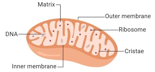 Mitochondria
Function:
- Break down glucose and other molecules during cellular respiration
- Energy released is stored as ATP (Adenosine Triphosphate) – the energy currency of the cell
- ATP is used to power almost all cellular activities
Why many small mitochondria instead of one giant one? Many small mitochondria have a much greater total surface area than one large one – more surface area means more space for chemical reactions and faster energy production.
Special feature: Mitochondria have their own DNA and ribosomes – suggesting they were once free-living bacteria that got incorporated into larger cells during evolution.
G. Plastids – Centre for Food Synthesis in Plant Cells
Plastids are organelles found only in plant cells (and some algae). They are used for food synthesis and storage.
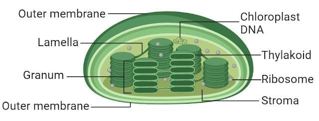
Three types of plastids:
| Type | Pigment/Content | Function | Example |
|---|---|---|---|
| Chloroplasts | Green pigment – chlorophyll | Photosynthesis – makes food using sunlight | Leaves |
| Chromoplasts | Yellow, orange, or red pigments | Give bright colours to flowers and fruits; attract pollinators and seed-dispersing animals | Flower petals, fruits |
| Leucoplasts | No pigment – colourless | Store food materials like starch, oils, or proteins | Potato (stores starch), taro/Colocasia |
Structure of Chloroplast:
- Double-membrane bound organelle (like mitochondria)
- Contains a semi-fluid substance called stroma
- Within stroma are disc-shaped membrane structures containing chlorophyll
- Light energy is absorbed during photosynthesis; sugars and starch are stored in stroma
Similarity with Mitochondria:Both mitochondria and plastids have their own DNA and ribosomes – this suggests they share an evolutionary history with ancient bacteria.
H. Vacuoles – The Organelles for Storage and Support
In plant cells:
- Usually one large central vacuole surrounded by a single selectively permeable membrane
- Filled with a watery fluid called cell sap
- Stores water, minerals, sugars, and waste materials
- By storing large amounts of water, the vacuole maintains pressure inside the cell → keeps the plant firm and upright
- When a plant does not get enough water → vacuole loses water → cells become less firm → plant wilts
In animal cells:
- Vacuoles are present but much smaller
- Help in temporary storage of materials
6. Prokaryotic vs Eukaryotic – Cell Organelle Comparison
| Structure | Bacterial Cell (Prokaryotic) | Plant Cell (Eukaryotic) | Animal Cell (Eukaryotic) |
|---|---|---|---|
| Cell membrane | Present | Present | Present |
| Cell wall | Present | Present | Absent |
| Cytoplasm | Present | Present | Present |
| Well-defined nucleus | Absent | Present | Present |
| Nucleoid | Present | Absent | Absent |
| Membrane-bound organelles | Absent | Present | Present |
| Chloroplast | Absent | Present | Absent |
| Mitochondria | Absent | Present | Present |
| Golgi apparatus | Absent | Present | Present |
| Lysosomes | Absent | Rarely present | Present |
| Large central vacuole | Absent | Present | Absent (small vacuoles) |
7. How Do Normal Cells Grow and Divide?
When you get a cut on your skin, it heals in a few days. When hair falls out, new hair grows. This happens because cells in our body can grow and divide to replace old, dead, or damaged cells.
Cells grow only up to a certain size and then divide to form new cells – this is how our body grows.
Cell division is the process by which new cells are formed from pre-existing cells. It allows organisms to:
- Grow
- Repair damaged tissues
- Reproduce
Both prokaryotic and eukaryotic cells divide, but eukaryotic cells divide in a more controlled and orderly manner through a process called the cell cycle.
Every day, an estimated hundreds of billions of cells in our body are replaced – about 1% of the total number of cells in the body.
7.1 Types of Cell Division
There are two major types of cell division:
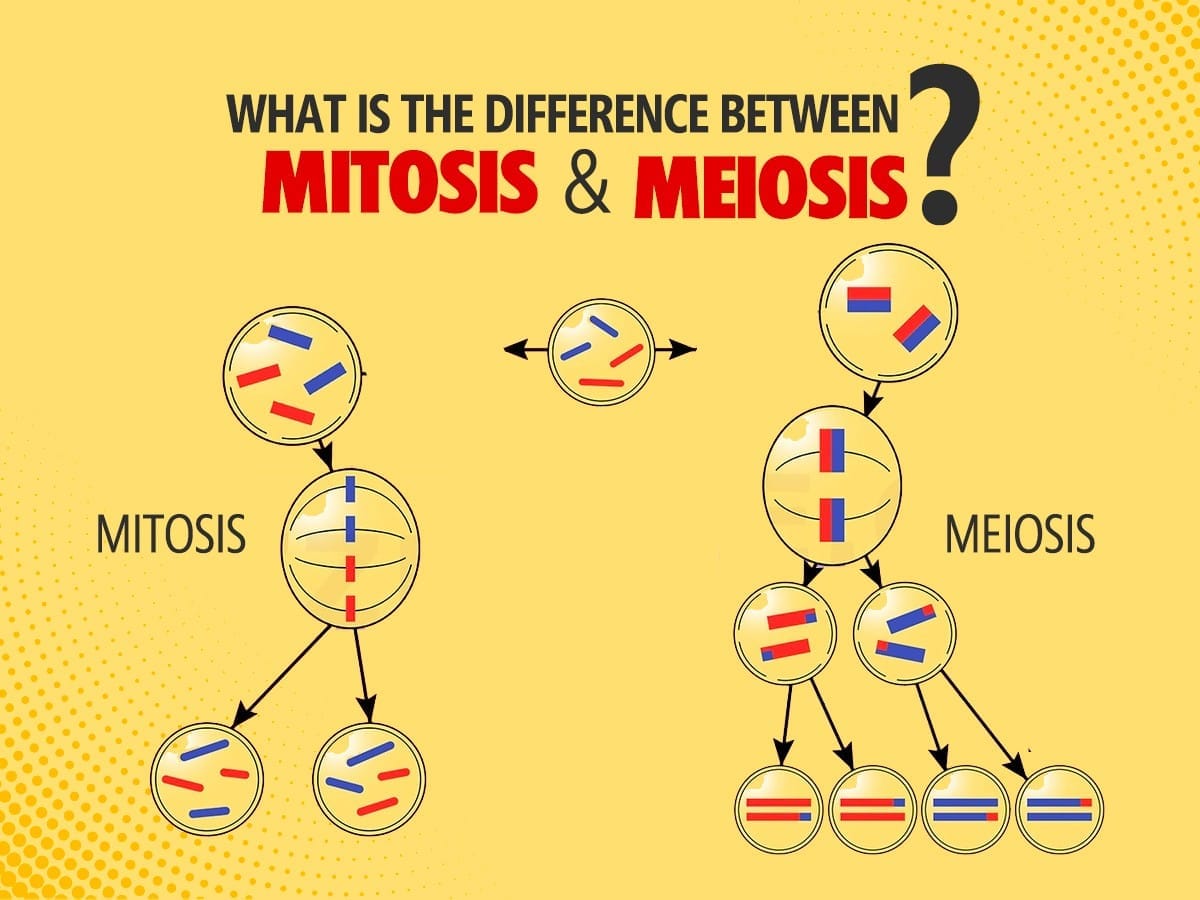
| Feature | Mitosis | Meiosis |
|---|---|---|
| Number of daughter cells produced | 2 | 4 |
| Chromosomes in daughter cells | Same as parent cell (full set) | Half the number of parent cell |
| Daughter cells identical to parent? | Yes – genetically identical | No – genetically different |
| Where it occurs | All body (somatic) cells | Reproductive organs only (testes, ovaries, anthers, ovaries in plants) |
| Purpose | Growth, repair, maintenance, asexual reproduction | Sexual reproduction; creates genetic diversity |
Mitosis in Simple Terms:
- One parent cell → divides → two genetically identical daughter cells
- Each daughter cell gets the same DNA and the same number of chromosomes as the parent
- This ensures genetic information is maintained across all body cells
- Every human begins as a single fertilised egg, which then undergoes mitosis trillions of times to form all the cells of the body
Meiosis in Simple Terms:
- One parent cell → divides twice → four daughter cells, each with half the number of chromosomes
- Occurs only in reproductive organs
- In humans: meiosis in testes (males) produces sperm; meiosis in ovaries (females) produces eggs
- In plants: meiosis in anthers (male parts) forms pollen; in ovaries (female parts) forms egg cells
- When sperm and egg combine during fertilisation → original chromosome number is restored
- Because meiosis creates variation, children resemble but are not exactly like their parents
What happens if cell division goes wrong?
- Errors in mitosis → uncontrolled cell divisions → tumours and abnormal number of chromosomes
- Errors in meiosis → genetic disorders, developmental problems, reduced fertility
8. Cell Theory – The Unifying Principle of Biology
History of Cell Theory:
| Scientist | Year | Contribution |
|---|---|---|
| Robert Hooke | 1665 | First observed cells in cork |
| Matthias Schleiden (German botanist) | 1838 | All plants are made up of cells |
| Theodor Schwann (German zoologist) | 1839 | All animals are made up of cells |
| Rudolf Virchow (German scientist) | 1855 | New cells arise only from pre-existing cells |
The Classical Cell Theory states:
- All living organisms are made up of one or more cells
- The cell is the basic unit of structure and function in living beings
- All cells arise from pre-existing cells
This unifies all of biology – from bacteria to humans – and explains life’s continuity through cell division.
Do Cells Grow and Reproduce Forever?
No. Cells grow and divide in a controlled way. They eventually die when they are no longer needed, and are replaced by new cells. Every cell has a definite lifespan.
Contact Inhibition: In many animal cells, cell division stops when cells come into contact with neighbouring cells. This is called contact inhibition – a natural “stop signal.”
Cancer cells lose this control and keep dividing uncontrollably → formation of tumours.
- Benign tumours – stay in one place
- Malignant tumours – can invade nearby tissues and spread to other parts of the body
Programmed Cell Death (PCD): Cells also have natural ways of dying in a controlled, genetically regulated manner. This is essential for normal development. For example, when an embryo forms fingers, PCD eliminates the cells between the digits – without this, we would have webbed hands.
Plant cells do not show contact inhibition because of their rigid cell walls – they follow a different pattern of growth.
9. Scientists Spotlight
Camillo Golgi: Italian scientist who in 1898 first observed the Golgi apparatus in nerve cells of a barn owl. Early microscopes could not resolve it clearly and many doubted its existence. Electron microscopy confirmed it decades later. It was named the ‘Golgi apparatus’ in his honour.
Arun Kumar Sharma: Famous Indian scientist known for his work on chromosomes, plant taxonomy, evolution, and development. He invented useful lab methods to study chromosomes in plants and received the Shanti Swarup Bhatnagar award and Padma Bhushan.
Gottlieb Haberlandt (Austrian botanist, 1902) proposed that any living plant cell, even from a permanent tissue, can develop into a complete plant if given suitable nutrients and conditions. This ability is called totipotency. His idea laid the foundation for Plant Tissue Culture Technology.
Quick Revision – Key Terms Table
| Term | What it Means |
|---|---|
| Cell | Basic structural and functional unit of all living organisms |
| Unicellular | Organism made of a single cell (e.g., bacteria, amoeba) |
| Multicellular | Organism made of many cells (e.g., humans, plants) |
| Limit of resolution | The minimum distance between two points that can be seen as separate – 0.1 mm for human eye |
| Cell membrane / Plasma membrane | Thin, selectively permeable outer boundary of all cells |
| Selectively permeable | Allows only certain substances to pass through |
| Osmosis | Movement of water through a selectively permeable membrane from dilute to concentrated solution |
| Diffusion | Movement of particles from higher to lower concentration (no membrane needed) |
| Hypotonic solution | Outside has less solute than inside the cell – water enters the cell |
| Hypertonic solution | Outside has more solute than inside the cell – water leaves the cell |
| Isotonic solution | Equal solute concentration on both sides – no net water movement |
| Cell wall | Rigid outer covering of plant, fungal, and bacterial cells – made of cellulose in plants |
| Fluid Mosaic Model | Model describing cell membrane as a flexible lipid bilayer with proteins embedded in it |
| Prokaryotic cell | Cell without a well-defined nucleus (e.g., bacteria) |
| Eukaryotic cell | Cell with a well-defined, membrane-bound nucleus (e.g., plant and animal cells) |
| Nucleus | Control centre of the cell – contains DNA and chromosomes |
| Nucleoid | Region in prokaryotic cells where circular DNA is located (no membrane around it) |
| Chromatin | Loosely arranged DNA + protein in non-dividing cells |
| Chromosomes | Condensed, rod-shaped structures of DNA + protein visible when cell is dividing |
| Genes | Functional segments of DNA that carry genetic information |
| Nucleolus | Dense body inside nucleus – site of ribosomal subunit synthesis |
| Ribosomes | Site of protein synthesis; present in both prokaryotic and eukaryotic cells |
| Endoplasmic Reticulum (ER) | Network organelle for synthesis and transport of proteins and lipids |
| RER | Rough ER – has ribosomes; makes proteins |
| SER | Smooth ER – no ribosomes; makes lipids and hormones |
| Golgi apparatus | Packages and ships proteins/lipids; post office of the cell |
| Lysosomes | Clean-up organelle – breaks down waste and damaged cell parts using enzymes |
| Mitochondria | Powerhouse of the cell – produces ATP through cellular respiration |
| Cristae | Finger-like folds of inner mitochondrial membrane – increase surface area |
| ATP | Adenosine Triphosphate – the energy currency of the cell |
| Plastids | Organelles found only in plant cells – for food synthesis and storage |
| Chloroplasts | Green plastids – perform photosynthesis |
| Chromoplasts | Coloured plastids (yellow/orange/red) – give colour to flowers and fruits |
| Leucoplasts | Colourless plastids – store food like starch, oils, proteins |
| Chlorophyll | Green pigment in chloroplasts that absorbs light for photosynthesis |
| Stroma | Semi-fluid matrix inside chloroplast where sugars are stored |
| Vacuole | Storage organelle – large in plant cells, small in animal cells |
| Cell sap | Watery fluid in plant cell vacuole |
| Turgid | Cell swollen with water (plants in hypotonic solution) |
| Flaccid / Wilted | Cell lacking water (plants in hypertonic solution or drought) |
| Cell division | Process by which new cells form from pre-existing cells |
| Mitosis | Cell division producing 2 identical daughter cells – for growth and repair |
| Meiosis | Cell division producing 4 daughter cells with half chromosomes – for sexual reproduction |
| Cell cycle | Controlled, orderly process of eukaryotic cell division |
| Contact inhibition | Normal cells stop dividing when they touch neighbouring cells |
| Tumour | Mass formed by uncontrolled cell division (cancer cells lack contact inhibition) |
| Programmed Cell Death (PCD) | Controlled, genetically regulated process of cell death – essential for development |
| Cell Theory | All organisms made of cells; cell is basic unit; new cells from pre-existing cells |
| Totipotency | Ability of a plant cell to develop into a complete plant – basis of tissue culture |